The presently available knowledge of the caeruloplasmin structure provides a glimpse of its plasticity and the multitude of binding sites allude to elaborate mechanisms of multifunctional activity. 1 Although "one gene, one protein, one function" has been a paradigm of biochemistry, caeruloplasmin joins the escalating number of enlisted "moonlighting" proteins, 2 which present scientists with the challenge of identifying when, how and why they exert their multiple roles.3 The following have all been plausibly proposed as physiological functions of caeruloplasmin.4:
The principal function attributed to caeruloplasmin is that of the plasma ferroxidase activity. Caeruloplasmin adopts an important, regulatory role in iron metabolism whereby it assists the release of iron from cells prior to its uptake by transferrin, the protein central to iron metabolism.5, 6
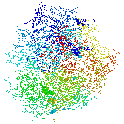
In caeruloplasmin the Type 1 Cu sites are buried at the bottom of a long, narrow and negatively charged crevice, beneath the divalent metal ion binding site.7 Situated above this, there is a large, more loosely ordered negatively charged patch that is believed to be the trivalent ion binding site. These Fe(III) binding sites may function in short range Fe(II) transport, shuttling up to four Fe(III) atoms between the putative membrane-bound Fe(II) donor and the ultimate Fe(III) acceptor in transferrin.
The oxidation reaction: Fe2+ --> Fe3+ + e- removes Fe(II) from the blood which may otherwise become involved in the generation of harmful reactive oxygen species.
Substantiation for caeruloplasmin ferroxidase activity comes from the identification of a mutation in the caeruloplasmin gene which is associated with systematic haemosiderosis (iron overload) in humans.8
Ascorbic acid (vitamin C) can be oxidised by one-electron steps to dehydroascorbic acid, by the way of two ascorbyl radicals.9 Ascorbate free radicals may be generated from ascorbic acid by reaction with superoxide and hydroxyl radicals; however, oxidation in the presence of transition metals, especially copper, is much more efficient. This oxidation is mediated by a catalytic redox system formed from the copper atoms bound to caeruloplasmin and iron atoms complexed by albumin.
Oxygen is directly reduced in water by addition of four electrons in one step, a process which circumvents the production of excited oxygen species as intermediates:
Concerted redox reactions occur between caeruloplasmin-bound Cu, albumin-bound Fe and ascorbate reducing Fe(III) and oxygen. The ferroxidase activity of caeruloplasmin also plays and integral rolein this redox system:
Caeruloplasmin is the principle mode of copper storage in the body; it binds an estimated 95 % of plasma copper whilst much of the remainder is bound in albumin.
Evidence for the role of caeruloplasmin in copper transport and storage has come from studies involving intravenous injection of radio-labelled copper and the association of caeruloplasmin with two hereditary copper transport disorders, Wilson`s disease and Menkes Syndrome. Interest has also been elicited by numerous reports on the existence of caeruloplasmin receptors in a variety of cell types,10 and such investigations have disclosed the peculiar ability of this protein to donate its copper to recipient tissue systems.
The discovery that copper metabolism is normal in patients with abnormal caeruloplasmin production, acaeruloplasminaemia, unquestionably demonstrates that caeruloplasmin has an essential role in iron, but not copper, metabolism.11,12
The blue copper oxidases couple the oxidation of a reducing organic substrate to the four-electron reduction of dioxygen to water. In the case of caeruloplasmin, an extensive group of organic substrates, including amino-phenols, p-phenylenediamines and catechols, can be oxidised in addition to ascorbate as previously mentioned.13 The oxidation of p-phenylenediamine constitutes a classic assay for the enzymatic activity of caeruloplasmin.14 This latter function is important since the random oxidation of amines would release nitrosoamines and other dangerously reactive entities into the blood plasma; amine oxidases help to remove excess transmitters or hormones such as adrenaline, which also requires Cu for its production in the vesicles of the adrenal gland.
Many biological substances, such as α-tocopherol and ascorbic acid, demonstrate both antioxidant and pro-oxidant activities, depending on the specific conditions. caeruloplasmin also falls into this category.
Chromatographic studies of human serum led to the proposal that caeruloplasmin is the primary antioxidant and barrier against free radicals in the bloodstream. Copious mechanisms have been proposed for caeruloplasmin antioxidant activity, including the sequestration of free copper ions and scavenging of O2·-, although most evidence suggests caeruloplasmin ferroxidase activity is of greatest importance; conversion of Fe(II) to Fe(III) may reduce oxidation by inhibition of the Fenton reaction, 15 which requires the reduced metal, by decreasing the amount of pro-oxidant Fe(II) available. Purified human caeruloplasmin has been shown to inhibit the oxidation of tissue extracts of lipids, polyunsaturated fatty acids and phospholipids. caeruloplasmin antioxidant activity also blocks protein and DNA damage and removes reactive intermediates that, in the case of hydrogen peroxide,16 17 a membrane-permeable oxidant, could lead to an altered intracellular redox state of vascular cells.
The same ferroxidase activity of caeruloplasmin that elicits antioxidant activity causes oxidative damage to macromolecules, in particular low density lipoproteins, under well-defined experimental conditions. During investigation into this process, extensive dialysis of caeruloplasmin did not alter its pro-oxidant activity, indicating the pro-oxidant copper was tightly bound. However, on treatment with the solid phase chelating ligand, Chelex-100, caeruloplasmin pro-oxidant activity was completely inhibited.18 The postulation that the seventh, labile surface copper atom is solely responsible for the pro-oxidation of LDLs is therefore corroborated. The most buried residue, His426, is also essential to the function of the pro-oxidant site which, once reduced, can be directly reoxidised by oxygen, giving rise to ROS production.
Oxidation of LDL in blood vessel walls may well be a collateral process with unfortunate consequences since it leads to the progression of atherosclerotic disease. Although much has been learned about cellular mechanisms of LDL oxidation by caeruloplasmin, the role of O2-· in the initiation of LDL oxidation is a proposal yet to be substantiated.19
Nitric oxide is a new entry to the list of caeruloplasmin substrates and the role for caeruloplasmin in the metabolism of nitrosothiols has been recently confirmed by clinical studies.20 Nitrosothiol adducts of nitric oxide function as nitric oxide transporters and are involved in the regulation of inter- and intra-cellular signal transduction.21 Nitric oxide appears unique among the substrates of caeruloplasmin since it is able to reach and directly bind to the blue copper sites.22 Nitric Oxide released by cells binds to Type 1 copper and is oxidised to nitrosonium, NO+, which then reacts with thiols at an enhanced rate.
1.Musci, G., Bellenchi, G.C. and Calabrese, L. Eur. J. Biochem., (1999) 265, 589 – 597.
2.Jeffery, C.J. Trends Biochem. Sci., (1999) 24, 8 – 11.
3.Bielli, P. and Calabrese, L. Cell. Mol. Life Sci., (2002) 59, 1413 – 1427.
4.Frieden, E. and Hsieh, H.S. Adv. Enzymol., (1976) 44, 187-236.
5.Osaki, S., Johnson, D.A. and Frieden, E. J. Biol. Chem., (1966) 241, 2746 – 2751.
6.Osaki, S., Johnson ,D.A. and Frieden, E. J. Biol. Chem., (1971) 246, 3018 – 3026.
7.Lindley, P., Card , G., Zaitseva, I., Zeitsev, V., Reinhammer, B., Selin-Lindgren, E. et. al. J. Biol. Inorg. Chem., (1997) 2, 454 – 463.
8.Harris, Z.L., Durley, A.P., Man, T.K. and Gitlin, J.D. Med. Sci., (1999) 96, 10812-10817.
9.Mouithys-Mickalad, A., Deby, C., Deby-Depont, C. and Lam, M. Biometals (1998) 11, 81-88.
10.Harris, E.D. Proc. Soc. Exp. Biol. Med., (1991) 196, 130 - 140.
11.Morita, H., Ikeda, S., Yamamoto, K., Morita, S., Yoshida, K., Nomoto et. al. Ann. Neurol., (1995) 37, 64 – 65.
12.Spira-Solomon, D.J., Allendorf , M.D. and Solomon E.I. J. Am. Chem. Soc., (1986) 108, 5318 – 5328.
13.Frieden, E. and Hsieh, H.S. Adv. Enzymol., (1976) 44, 187 – 236.
14.Musci, G., Bellenchi, G.C., and Calabrese, L. Eur. J. Biochem., (1999) 265, 589-590.
15.Burkitt, M.J. Prog. Reac. Kin. Mech., (2003) 28, 75-103.
16.Cha, M.K. and Kim, I.H. Biochemistry, (1999) 38, 12104 – 12110.
17.Park, Y.S., Suzuki, K., Taniguchi, N. and Gutteridge, J.M.C. F.E.B.S. Lett., (1999) 458, 13-136.
18.Mukhopadhyay, C.K., Mazumder, B., Lindley, P. and Fox, PL. Proc. Natl. Acad. Sci., USA (1997) 94, 11546-11551.
19.Mukhopadhyay, C.K.and Fox, P.L. Biochemistry, (1998) 37, 14222 – 14229.
20.Moriel, P., Pereira, I.R., Bertolami, M.C. and Abdalla, D.S. Free Radic. Biol. Med., (2001) 30, 318 – 326.
21Akaike, T. Free Radic. Res., (2000) 33, 461 – 469.
22.Musci, G., Di Marco, S., Bonaccorsi di Patti ,M.C. and Calabrese, L. Biochemistry, (1991) 30, 9866 – 9872.