Ischæmia - story in pictures!
Evidence that free radicals can be formed in vivo in blood during a variety of medical procedures has been accumulated over a number of years. One area of medical concern is that free radicals can be formed when blood is reperfused into a part of the body which has been previously ischaemic. This is especially important if it occurs in the heart as it can possibly lead to heart attacks. We were approached by a heart surgeon conducting angioplasty on his patients to see if we could investigate reports of a "radical burst" following such angioplasty.
Free radicals have been trapped using spin trapping techniques from blood samples removed through a catheter. In the present study using a dozen healthy volunteers in the age range 20-40, forearm ischæmia was induced by cuffing with a tourniquet for 4 minutes. reperfusion then followed after the tourniquet was released. Blood samples were taken before and after the arm was made ischæmic, then subjected to spin trapping using PBN, followed by EMR measurement of toluene extracts of the products.
A signal "burst" occured in most of the experiments, but this was very unpredictable when compared to the experiments performed on animals. However the animal experiments all involve very extreme ischæmia, often with the removal of organs, and invariably with the death of the unfortunate subjects. We were not allowed to sacrifice our student volunteers - though a couple of them did faint on seeing the needle!
During the experiments we noticed an unusual set of signals in many of the toluene/PBN extracts. This story concerns attempts to identify those signals.
Michael Bamford, Matthew Barker, Colin Bolton, Sarah Crossley, Jeremy Dwight, Martin Hartog, Stephen Koehring, John Maher, Jonathan Pinkney and thanks to Huayun Jackson
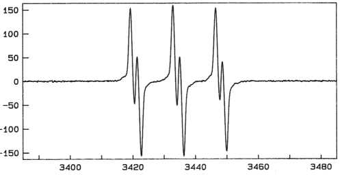
Normal PBN-trap signal observed
Abnormal PBN-trap signal observed
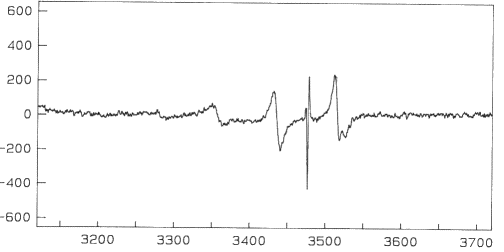
This one before applying the tourniquet
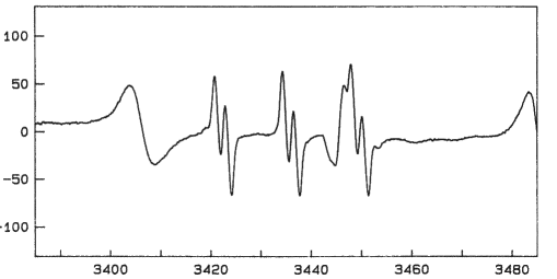
Note that the scale on the lhs has changed - the signal increased considerably - and what we now identify as a copper signal, disappears.
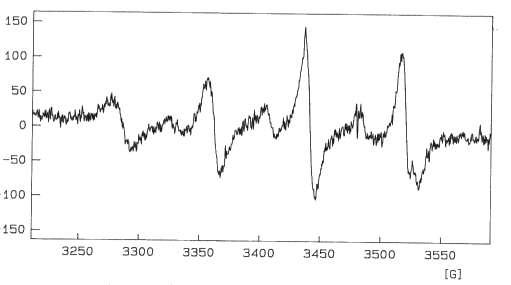
.. but this one after the tourniquet was released
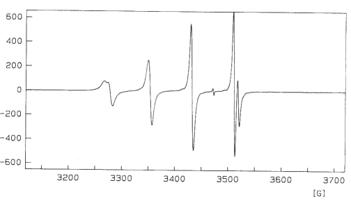
The whole signal is now shown, a quartet with an additional doublet splitting on the peaks

This is clearly a copper complex.
Note the Cu63/65 splitting on the high field component.
A weak signal from some PBN radical trap is still visible
A great similarity to BisDiethyldithiolatoCopper(II)
There are reports of such Cu(II) signals from rubber
Leaving a black rubber bung in contact with toluene, water, and some surfactant AOT
The AOT was to form micelles so that more water would enter the toluene. The sharp line comes from the micelles.
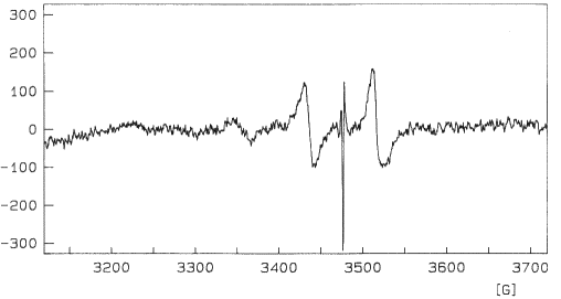
A similar test with one of the black caps from the storage tubes used in the tests
Serendipity! Signal from oxidative decomposition of a Cu(I)pyrazoylborate complex
 NB - this signal was disappearing rapidly, and originally looked
far more like that of the Cu(II) dithiolate above - despite numerous
attempts, we have never managed to repeat the observation.
NB - this signal was disappearing rapidly, and originally looked
far more like that of the Cu(II) dithiolate above - despite numerous
attempts, we have never managed to repeat the observation. A crystal structure of the Cu(I)trimer
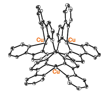
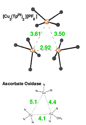
The Cu(I) trimer structure shows some similarity to that of ascorbate oxidase
Caeruloplasmin is the main source of copper in blood serum
The structure of a pure sample of caeruloplasmin has recently been measured by X-ray diffraction, it showed a copper trimer center, as well three other remote coppers.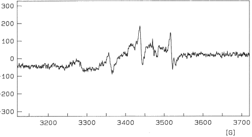
This shows clearly that hCp can generate the copper signal we observe.
Thanks to Professor Peter Lindley and Dr Slava Zaitsev of CCLRC Daresburyfor a pure sample of caeruloplasmin to carry out this test.Summary: Blood Concentration of Copper
The concentrations are of the order of 300mg dm-3, the 7ml of blood used in the tests would provide about 10-7 moles of copper, measurements showed that the signals we observed in some of the ischæmia tests could be achieved with 10% of the available copper. Moreover the copper could not have come from the apparatus. The observations of traces of copper in some rubber bungs and in the plastic caps of the centrifuge tubes, or in the cap of the Vacutainer tube used to hold the blood, were only obtained from prolonged contact with these implements. During the ischæmia tests contact times were very much shorter, and in most cases there was no contact between any of the blood extraction processes and any possible copper source.