Uses
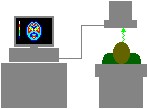
The main use of 99mTc is in diagnostic imaging. A freshly prepared sample of 99mTc is administered intravenously to the patient. After a short period the patient is placed under a gamma camera which detects the individual radioactive decays and where they originated.
There are a number of different radiation detection devices. The scintillation counter is by far the most widely used in nuclear medicine.
Scintillation counters are made up of a sodium iodide crystal coupled to a photomultiplier tube. When gamma rays strike the crystal, flashes of blue-violet are produced. The crystal is transparent to light and it’s enclosed in a light tight container. A powder is used to reflect light out, only through the crystal area adjacent to the photomultiplier tube. Once the flashes of light reach the surface of the photomultiplier tube, electrons are released.
These electrons become amplified in the photomultiplier tube and are amplified further. Now the electrons are ready to be processed and displayed. The light emitted from the output signal from the detector unit is proportional to the energy released by the gamma ray.
Collimators play a major part in detector systems. Collimators are designed so that the detector can only see photons in a specific area inside a patient while rejecting others from outside this area. The wide angle collimator is most commonly used and each type is designed for a specific purpose. When counts from a large field of view are needed the wide angle is used.
The electrical pulses of electrons are directed to the processing unit from the detector system. A spectrometer is used to sort the spectrum of gamma ray energy and accept or reject ones of specific energy. The window must be adjusted to reject all rays above and below certain energy levels. A window is the range of energy of an isotope. In the case of technetium, its energy range is 140keV. The energy range is set up for 20% of the isotope, one will allow 10% above and 10% below the energy range. This will help to reduce the counts from scattered radiation.
From the spectrometer the information is passed on to be displayed as counts on a scaler. A scaler measures the amount of radioactivity from within the source.
Scanners are designed to produce two-dimensional pictures of the distribution of the radioactive isotope in an organ. Below is an example of a full body scan.
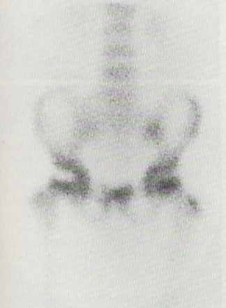
Please click on thumbnail for a larger image
Many other different types of imaging are possible