The
most common method of DNA typing is RFLP analyisis of VNTR loci.
Following
standard protocols DNA is extracted and purified. It is cut into fragments using
enzymes called restriction endonucleases, which recognize short DNA sequences
and cleave only at or near specific tandemly repeated base sequences. Such
areas, known as restriction enzyme recognition sites, occur on either side of
the polymorphic locus of interest. Since the DNA fragments are of variable
lengths, they are referred to as restriction fragment length polymorphisms (RFLPs).
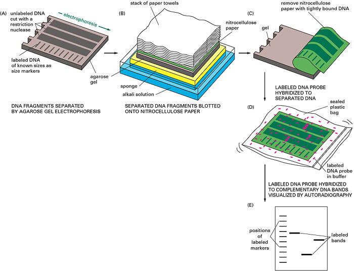
These fragments are sorted on the basis of size by electrophoresis in an agarose
gel. This involves applying an electric current through a gel with the current traveling
from the negative electrode at the top of the gel to the positive
electrode at the bottom of the gel. The DNA fragments are negatively charged due
to the phosphate groups and therefore travel toward the positive electrode. The
smallest fragments migrate to the bottom and the largest remain on top. DNA is
made single-stranded or denatured and a nylon membrane is placed on top of the
gel. DNA is then transferred to the membrane via capillary action and fixed by
baking making it accessible to a probe. A radioactive probe specifically binds
to appropriate VNTR fragments by complementary base pairing. X-ray film is
placed over the membrane and the DNA profile visualized by auto-radiography.
Step
A - Separation of nucleic acids band on size by agarose gel electrophoresis.
Step
B & C - Transfer of separated nucleic acids from gel to nitrocellulose
paper.
Step
D - Hybridization with radioactive probe.
Step
E - Auto-radiograph of DNA profile.
This process, known as the Southern Blot Technique, was developed in 1975 and adopted by Jeffreys et al for RFLP analysis for the first DNA fingerprinting in 1985. The DNA banding patterns were difficult to interpret as he used probes containing core sequences of base pairs that identified tandem repeats on many different chromosomes. The following year the single locus probe method was introduced becoming the preferred method.