
THE EYE

![]()
![]()
The below diagram shows the basic anatomy of the human eye:
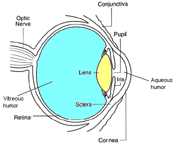
Cornea:
the transparent part at the front of the eye that refracts light entering
the eye onto the lens.
Iris:
This is the coloured part of the eye that controls the amount of light
that enters the eye, it is able to contact and dilate in order to control
the size of the pupil depending on the light intensity.
Sclera:
the outer white part of the eye that protects the inner structures.
Optic
nerve:
this leaves the eye at the optic disk and transfers all the visual
information to the brain.
Conjunctiva: a transparent vascular membrane that lines the inside of the eyelids and extends over the front of the white part of the eye (the sclera).
Aqueous
humor:
this fluid circulates the front part of the eye, it provides nourishment
and helps maintain the eye pressure.
Vitreous
humor:
the clear gel in the centre of the eye that helps the eye to maintain its
spherical shape.
![]()