
Photoreceptors are modified neurons.
Structurally they resemble tall epith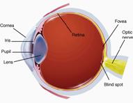 elial
cells turned upside down with their tips immersed in the pigmented layer of
the retina. Going from the pigmented layer into the neural layer,
rods and cones have an outer segment joined to an inner segment by a
stalk containing a cilium.
The inner segment connects to the cell body, or nuclear region, which is
continuous with an inner fibre tipped with synaptic endings.
elial
cells turned upside down with their tips immersed in the pigmented layer of
the retina. Going from the pigmented layer into the neural layer,
rods and cones have an outer segment joined to an inner segment by a
stalk containing a cilium.
The inner segment connects to the cell body, or nuclear region, which is
continuous with an inner fibre tipped with synaptic endings.
Image showing the position of the retina in the eye. Taken from http://cwx.prenhall.com/bookbind/pubbooks/morris2/ chapter3/medialib/summary/1.html, without permission.
The light absorbing visual pigments are packaged in membrane bound discs within the outer segments. The coupling of the photoreceptor pigments to cellular membranes magnifies the surface area available for light trapping. In rods the discs are discontinuous and stacked like hollow pennies within the plasma membrane. In cones the discs become increasingly smaller towards the end of the cell and their membranes are continuous with the plasma membrane. Photoreceptor cells are vulnerable to damage. If the the retina becomes detached, the photoreceptors begin to degenerate. They are easily destroyed by intense light.
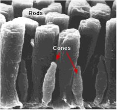
This scanning electron micrograph (courtesy of Scott Mittman and David R. Copenhagen) shows rods and cones in the retina of the tiger salamander.Taken from http://users.rcn.com/jkimball.ma.ultranet/BiologyPages/V/Vision.html without permission
The rods and each of the three cone types contain unique visual pigments, that absorb different wavelengths of light and have different thresholds for activation. Visible wavelengths of light are picked up allowing us to visualise the spectrum of light shown below.
Taken from http://cwx.prenhall.com/bookbind/pubbooks/morris2/chapter3/ medialib/summary/1.html without permission.
Rods are very sensitive and even respond to dim light making them best suited for night vision, however their inputs are only perceived as grey tones. Cones require very bright light for activation but allow us to view the world in an array of vivid colours. Rods and cones are attached differently to other retinal neurons causing further differences in their abilities. Rods are in converging pathways and up to 100 rods may feed into a single ganglion cell. Due to this, rod effects are considered collectively resulting in vision with poor resolution. In contrast each cone fovea has a direct pathway to a single ganglion cell. This accounts for the sharp detailed high resolution views of very small areas of the visual field provided by cones.