 |
 |
 |
 |

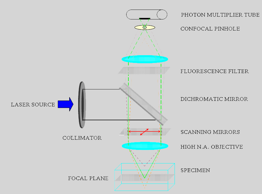
Why Confocal Microscopy? - When using a conventional microscope light from the sample is collimated by the lens and focused to the eye. However, there is a limit where light from out of the plane of the object enters the eyepiece - this out of plane light causes the perceived image to be blurred. By using this technique the out of plane light is prevented from entering the eyepiece thus eliminating the blurred effect.
The principal idea was first proposed in the late 1800‘s by a German engineer, Nipkow. The technique was pioneered by Minsky (a Harvard postdoc) in the 1950‘s. He built and patented a ‘double focusing stage scanning microscope‘[1,2] in 1957.
A point source of light is created using the light source A through pinhole B. The objective lens C is used to focus the light to a diffraction limited spot D within the specimen. The second objective lens is used to collimate the light from D onto the exit pinhole F to be detected by a phototube G.
B, D, and F are at optically conjugate focal planes - hence the name confocal - coined by Brakenhoff [3] in the 1970‘s. Light that is not in the confocal plane will be focused either before or after the pinhole - thus light that is not in the confocal plane will not reach the detector; the out of focus blur associated with conventional full-field microscopy is prevented.

[1] Minsky, M.
"Microscope Apparatus"
U.S. Patent 3013467 (1961) Filed 1957
[2] Minsky, M.
"Memoir on inventing the confocal scanning microscope"
Scanning 10 (1988) p.128
[3] Brakenhoff, G.J. Bloom, P. and Barends, P.
"Confocal scanning microscopy with high-aperture lenses"
Journal of Microscopy 117 (1979) p.219