
Below is a schematic plan view of the ablation chamber. An excimer laser (Lambda Physik, Compex 201) charged with ArF (193 nm) with a typical pulse energy of 2/300 mJ is focused using a fused silica lens (focal length = 27 cm) onto the target mounted in a stainless steel vacuum chamber maintained at ~10-6 Torr. The area of the focal volume has been estimated by stylus profilometry to be ~ 1 x 0.4 mm2.
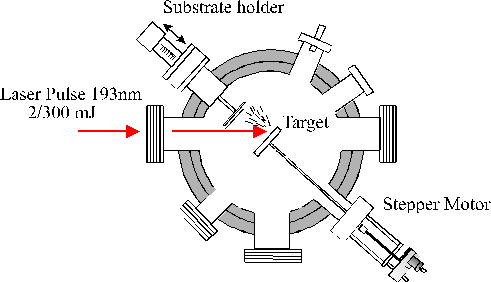
The target takes the form of a 50 mm disk mounted on a shaft attached via a double 'O' ring seal to a stepper motor. This enables the target to be rotated during ablation to prevent deep crater formation, allowing longer deposition times and reducing the number of large particulates being sputtered from the target and being incorporated into the films. In earlier experiments the target took the form of a cylindrical rod suspended vertically from the lid of the vacuum chamber, and translated and rotated via a stepper motor. The shot-to-shot reproducibility of such an arrangement is extremely sensitive to any eccentricity in the rod rotation. Thus the present set-up employs a side mounted rotating disk with the laser normally incident at an angle of 45°.
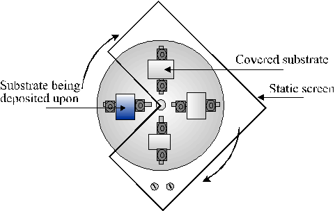
Films are deposited on various substrates, including single crystal silicon, quartz and NaCl, each ~1 cm2 in area. The substrate holder allows the target substrate distance to be varied between 30 and 60 mm by means of a micrometer and bellows arrangement. The substrate holder is mounted on a 360° rotatable flange, which allows up to four film to be deposited independently by rotating the samples behind a static screen.
| The focal volume adjacent to the target, and the ablation plume, are both clearly visible via their accompanying optical emission. The light emitted from the expanding plume is imaged through a lens onto a quartz fibre optic bundle (Oriel) located behind a lens/iris combination and an appropriately positioned quartz observation port mounted in the top flange of the chamber. This optical arrangement restricts the viewing zone in the vicinity of the plume to a column estimated at ~2 mm in diameter and is translatable to provide spatial resolved emission spectra. An example of the expanding emission plume can be seen on the right. |  |
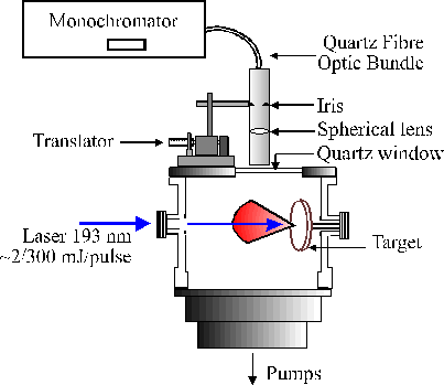 The other end of the fibre bundle butts up against the entrance slit of either of two monochromators. One is a 12.5 cm monochromator (Oriel), equipped with a 600 lines/mm ruled grating and an Instaspec IV CCD array detector, which is capable of providing low resolution dispersed emission spectra covering a wide (~300 nm) range of wavelengths from just a single laser shot, the signal to noise of which can be enhanced by summing for many laser shots. The second is a 0.5 m Spex 1870 monochromator equipped with a 2400 lines/mm holographic grating and entrance and exit slits whose widths are user selectable. Light emerging from the exit slit of this monochromator is detected with a red sensitive photomultiplier tube (PMT). Higher resolution emission spectra were obtained by scanning the monochromator with the slit widths set narrow and passing the PMT output via a boxcar and a V/F converter to a PC.
We have recently built a state-of-the-art QMS sampling system to allow us to study the species within the ablation plume. This system is a Hiden Analytical EPS QMS with energy analysis facility. The axis of the QMS is aligned so that particles within the ablation plume impinge upon the opening to the QMS and are then ionised and detected. We can detect atoms, neutrals, ions (positive and negative), and clusters, as well as measure the KE distribution of each. |
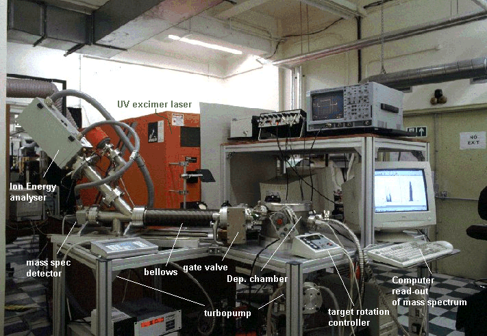 The Pulsed Laser Deposition apparatus and Mass Spectrometer sampling system (Click on photo for larger sized image, 342 kb) |
