Positron Emission Tomography
Fundamental Principles
A positron emitting isotope is introduced into the body
usually intravenously and the isotope accumulates in the target organ. The
positron quickly reacts with an electron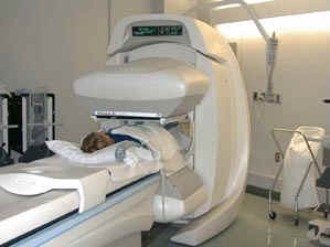 producing two gamma rays in opposite
directions as they collide. The emitted gamma rays are detected by the PET
camera which can pinpoint the location. Organ malfunction can be assessed
due to areas where the radioisotope is in low concentrations is called a cold
spot or where it is taken up in excess known as a hot spot. The organ can
also be monitored of a long period of time and display irregular patterns of
activity or usual rates of the isotope movement also indicating a malfunction
producing two gamma rays in opposite
directions as they collide. The emitted gamma rays are detected by the PET
camera which can pinpoint the location. Organ malfunction can be assessed
due to areas where the radioisotope is in low concentrations is called a cold
spot or where it is taken up in excess known as a hot spot. The organ can
also be monitored of a long period of time and display irregular patterns of
activity or usual rates of the isotope movement also indicating a malfunction
Benefits
PET imaging has many advantage over other techniques. Since the uptake
of a radiopharmaceutical is biochemically based and diseases cause a disruption
to the usual chemical processes, this technique offers the earliest possible
diagnosis of a disease even before the onset of symptoms. This could mean
the disease can be more easily cured or prevented from progressing
further. It also is the one of the best ways of monitoring the success of
a treatment and so any adjustments can be correspondingly made.
Since the test is painless and non-invasive it has much lower level of risk
than with other diagnostic tests. PET scanning is preferable to x-ray
diagnostic tests since it subjects the patient to five times less radiation and
it can be used for soft tissue as well as bones. It is also the most
cost-effective method since other techniques are inherently more expensive due
to their invasive nature.
Applications
PET scans can be used to assess blood flow to the brain, the function of the
heart, liver and kidneys as well as checking bone development. The
radioisotope used must produce gamma rays of high enough energy in order for
them to be detected and have a short half-life so as to minimise the radiation
experienced by the patient after the scanning has finished. This website
will only go into more detail on brain and heart imaging.
►CONTINUE
www.snm.org/nuclear/benefits.html
www.uic.com.au/nip26.htm
http://neuro.pet.rh.dk/research/illustrations
www.crump.ucla.edu/software/lpp/pet_overview.html