
Interactive Raman spectra of adamantane, diamantane and diamond, and their relevance to diamond film deposition
PhysChemComm, 1998, 1, 35 - 44
DOI:
10.1039/a808830f
P. W. May,*a S. H. Ashworth,a C. D. O. Pickard,b M. N. R. Ashfold,a T. Peakmana and J. W. Steedsb
a School of Chemistry, University
of Bristol Bristol ,
UK BS8
1TS
b Department of
Physics, University
of Bristol Tyndall
Avenue , Bristol , UK
BS8 1TR
Received 11th November,
1998 , Accepted 18th
November, 1998
Laser Raman spectra have been obtained for crystalline adamantane, diamantane, and natural type II diamond, as well as for a diamond film grown by chemical vapour deposition (CVD). The spectra are presented in JCAMP format to allow on-line interaction with the data by the reader, and 3D structure animations are presented showing the most probable vibrational mode causing each peak in the various spectra. The spectra are compared to those often seen from CVD diamond films, and which contain peaks that have previously been assigned to 'nanophase' diamond. Although there is a superficial correlation between the peak positions for the polymantanes and those seen in CVD diamond, the conclusion is that the nanophase diamond peaks are not an intrinsic vibrational mode of a polymantane, but instead must be a result of some other aspect of the nanophase scale of the films, such as surface modes.
Link to Software requirements
Introduction
Laser Raman spectroscopy (LRS) is an important technique used to analyse solid state materials.1 Recently it has been extensively utilised to determine the quality of diamond films deposited by chemical vapour deposition (CVD),2-7 since the ratio of the characteristic sharp diamond peak at 1332 cm-1 to that of the broad feature at ~ 1550 cm-1 corresponding to the graphitic G-band is an indication of the phase purity of the sample.2 In some LRS studies of poorer quality diamond or of the initial stages of diamond growth, several peaks around 1130-1230 cm-1 have been observed,8 and have been tentatively assigned to 'nanophase' diamond: a term used to describe a material consisting of carbon having short range sp3 crystal order surrounded by a matrix of less ordered material. However these peak assignments are still controversial and more data are needed to clarify the situation. Apart from the diamond peak, the major peaks seen in this wavelength range are 1130,9 1140 10 and 1149 cm-1,8 all assigned to nanophase diamond, 1467 8 and 1480 cm-1 9 assigned to a 'diamond precursor' or polyacetylene, and 1200-1210 cm-1, which has been seen in highly defective films grown by hot filament CVD,11 in highly B-doped diamond films,12 and at the substrate interface after heteroepitaxial growth of diamond on Si,13 again being assigned to nanophase diamond. Confocal Raman studies of heteroepitaxial diamond film growth on Pt substrates14 revealed peaks at 1230, 1640 and 1470-1490 cm-1 assigned to defective disordered diamond nanocrystals, 1400 and 1530 cm-1 assigned to disordered graphite, and 1500 cm-1 assigned to a distorted sp3-bonded carbon network. Very recently, Prawer et al.15 obtained the LRS spectrum of so called 'amorphous diamond' (a randomly arranged but fully sp3-bonded carbon network) and found that it was dominated by a large peak at around 1200 cm-1.
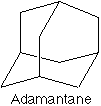
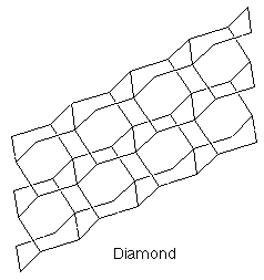
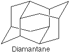
The nanophase diamond that allegedly gives rise to some of these peaks can be considered to be a collection of fused adamantane units, since adamantane, C10H14, is the smallest building block exhibiting the diamond structure (see Fig.1). Higher diamondoids in the series exist,16 continuing with diamantane, C14H20, triamantane, C18H24, and so on. The first three in this series have been studied using LRS and the resulting peaks assigned to either C-C or C-H vibrational modes.17,18 However these reports were performed before diamond CVD was established, and so spectra are not available in the literature which cover the wavenumber region of interest to diamond CVD researchers (although most of the peak positions are tabulated). This is partly the motivation for the present work: to use modern LRS systems to obtain the spectra of relevant diamondoid structures over an appropriate wavelength range, and to compare these results with features observed in diamond films. We have chosen to use the first two in the series, adamantane and diamantane, since these are readily commercially available.
Experimental
LRS was performed upon samples of adamantane, diamantane, natural diamond and a diamond film grown in house by CVD. Adamantane and diamantane (also known as congressane) were purchased in crystalline form from BDH and Aldrich Chemicals, respectively. The single crystal diamond sample was a 1 mm type IIa natural stone containing low levels of nitrogen and other impurities. The CVD diamond film was grown in a hot filament reactor using the standard conditions: 1% CH4 in H2, pressure 20 Torr, substrate temperature ~ 900 °C, Ta filament temperature ~ 2500°C for 8 h, using Si (100) as a substrate which had been manually abraded with 1-3 µm diamond grit prior to deposition. These conditions produced a 4 µm thick polycrystalline CVD diamond film, with random crystallite orientation.
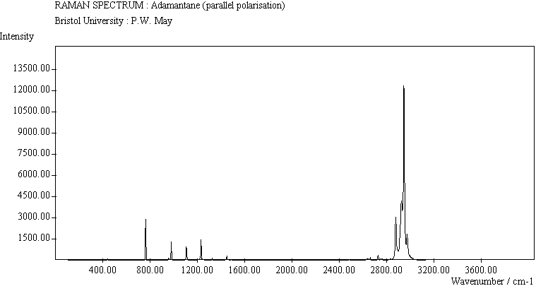
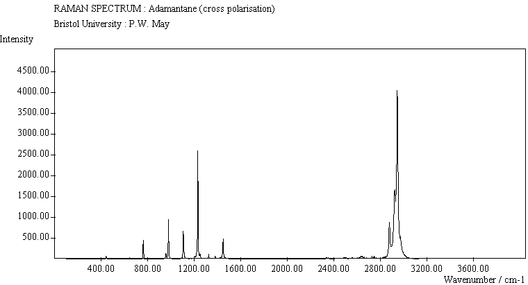
LRS was performed on a Renishaw System 1000 Raman operating with the 488 nm line of an Ar+ laser, in a backscattered configuration. The laser was linearly polarised, and a polarisation filter and quarter wave plate were used to give information about the symmetry of the vibrational modes in the samples. Spectra could be obtained with the filter parallel or perpendicular (crossed) to the laser polarisation.
The assignments were made on the basis of relatively low-level calculations carried out using the Gaussian 94 program suite (restricted Hartree-Fock calculations with 3-21G* basis.) The experimental Raman spectra were then matched to the overall patterns of the ab initio calculations and then modified in the light of the polarisation information. In general the calculated frequencies were over-estimated. The small unassigned peaks in the adamantane spectrum between 1500-2800 cm-1, and in the diamantane spectrum between 2250-2800 cm-1 are most probably combination bands. The animations of adamantane and diamantane have been produced using the optimized geometry and maximum displacements from the ab initio calculations. The individual frames have been calculated using a simple sinusoidal interpolation, and the amplitudes of the vibrations have been exaggerated in order to make them visible on the screen. The animation of the diamond lattice uses the fact that all the individual unit cells are in phase with one another (i.e. k=0). The diamond lattice can be thought of as two face-centred cubic lattices and in this case the two lattices as a whole are moving relative to one another. Again the animation uses a simple sinusoidal interpolation for each frame. The appropriate animated structure file is then linked to the correct wavenumber value (x-coordinate) of the JCAMP file by use of a Chime script.
Results
The various spectra are shown in Table 1 and Table 2. The JCAMP files (.jdx), shown in Table 1, are interactive files which can be viewed using a suitable reader, such as the Chime plug-in. This will allow the reader to zoom in and out of the spectra, and to save the data as x,y coordinates to their own computer. Animated 3D structure files (Animation 1) linked to the interactive spectra are available for adamantane, diamantane and natural diamond. When a peak in the spectrum is clicked upon the 3D molecular structure will animate, illustrating the vibrational mode most likely to have caused the peak.
Animation 1 Links to animated 3D structure files and JCAMP spectra.
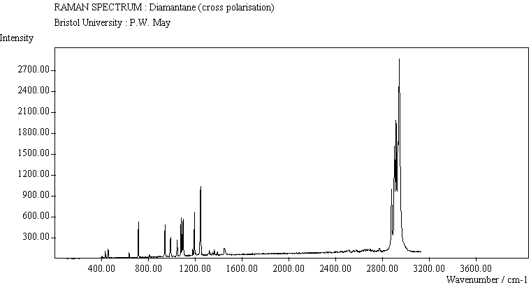
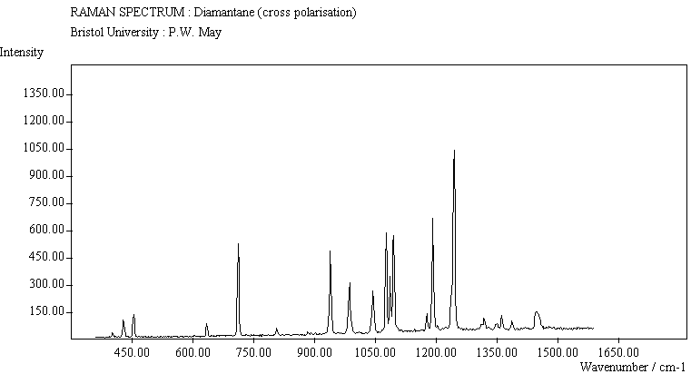
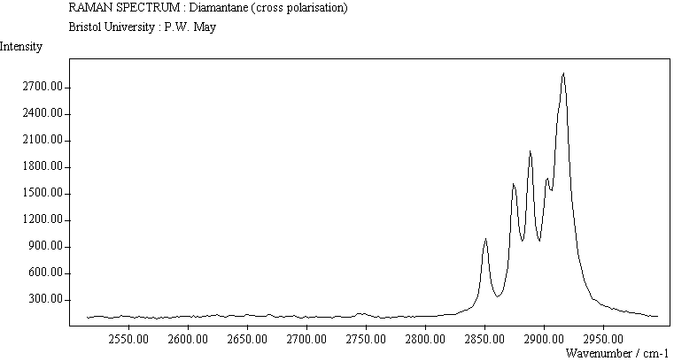
For readers without the appropriate software, the important parts of the spectra can instead be seen as GIF images in Table 2. These GIF images should also be used for hardcopy prints, if required.
Discussion
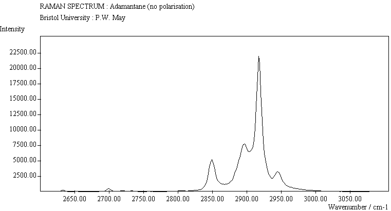
The spectra for the two polymantanes show two regions, one at low energy (roughly 300-1600 cm-1) corresponding to the various C-C skeletal bends, rocks and stretching modes, and one at high energy (~2600-3000 cm-1) corresponding to the C-H stretching modes. Using the 3N-6 rule for the number of vibrational degrees of freedom: adamantane has 24 atoms = 66 modes and diamantane has 34 atoms = 96 modes, although, of course, only some of these will be Raman active.
Adamantane and diamantane
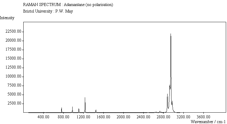
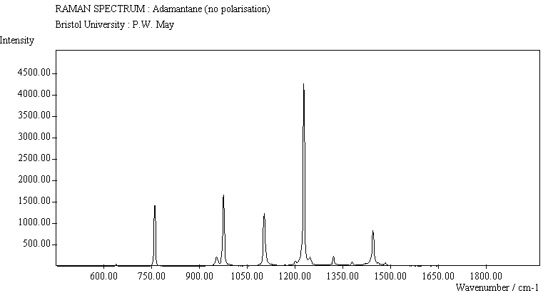
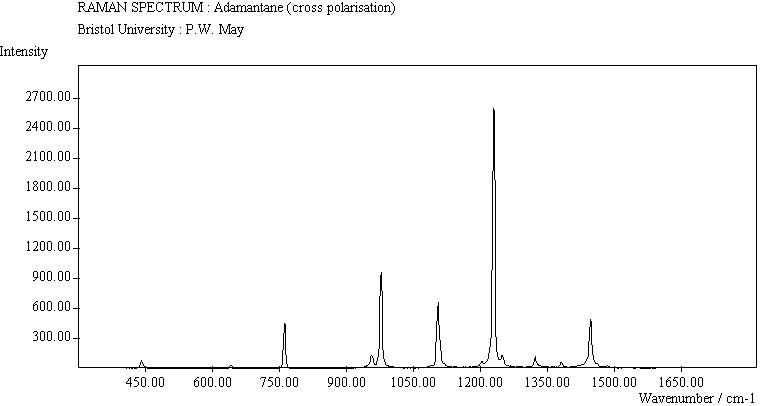
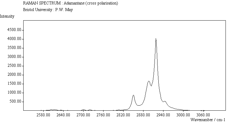
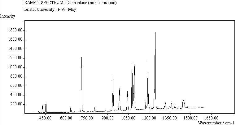
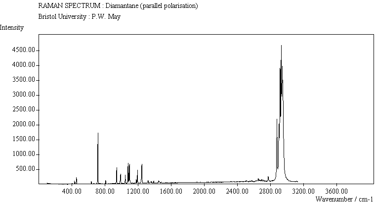
The peak assignments for adamantane and diamantane have been taken from ref.18 and are reproduced in Table 3. The peaks in our spectra agree with these values to within the resolution of the spectrometer (~1 cm-1). There are some differences, though, especially at lower energies. Our spectra reveal some lines which were only present at low temperature in ref. 18. This could be because we used a higher energy laser for the excitation, which might cause resonance effects, or that our detector was more sensitive at these lower energies. We have also seen a number of peaks between 1472-2804 cm-1 which have not been reported before and so have yet to be assigned. These peaks could be a result of second order scattering processes or combination bands. By comparing the relative heights of the various peaks to their neighbours for different polarisations, we can assign some of the peaks to the totally symmetric class A1. For adamantane, these occur at 758, 1097, 1312 and 2941 cm-1, whilst for diamantane these are at 453, 708, 802, 933, 1037, 1299, 1307, 1459, 1465, 2846, 2898 and 2920 cm-1.
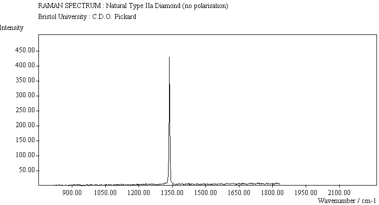
Single crystal diamond
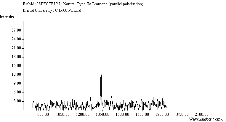
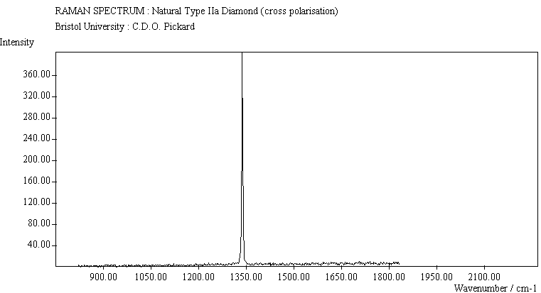
The diamond spectra show that only one sharp strongly polarised line is seen, at 1333 cm-1, characteristic of the diamond fully symmetric stretching mode.
CVD diamond film
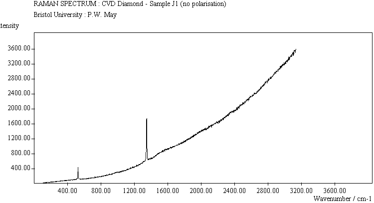
The spectra from the CVD diamond film show two peaks superimposed upon a strongly rising photoluminescence background. The peak at ~ 1334 cm-1 corresponds to the diamond peak, shifted slightly due to stress effects. The peak at 521 cm-1 is from the Si substrate, which interacts with the laser through the thin diamond coating.
Conclusions
The polymantane spectra exhibit many peaks in the wavenumber region where nanophase diamond peaks have been reported, and as the size of the diamondoid structure increases the number and complexity of the spectral lines also increase. There is some correlation between the peaks assigned to nanophase diamond and those seen for the two diamondoids, but the match is not convincing. Adamantane and diamantane have peaks around 1200-1250 cm-1, (at 1219 and 1234 cm-1, respectively), and again between 1430-1480 cm-1 (1434 and 1434 cm-1, respectively). But if these are the lines assigned to nanophase diamond, we would expect to see many of the other, stronger, diamondoid lines as well (such as peaks around 1030-1090 cm-1), rather than just one or two. This suggests that the spectral lines seen in diamond films are not due to the intrinsic vibrational modes of a diamondoid structure. This leads to the conclusion that some other aspect of the nanophase scale of the films is responsible for the observed features. One possibility is surface phonons and/or edge effects, which also have been found to have peak positions within the correct wavelength ranges.19
Acknowledgements
PWM thanks the Royal Society, and SHA thanks the EPSRC for personal funding.
The authors also wish to thank Renishaw
References
|
|
T. R. Gilson and P. J. Hendra, Laser Raman Spectroscopy,
Wiley-Interscience, |
|
|
|
D. S. Knight and W. B. White, J. Mater. Res., 1989, 4, 385. |
|
|
|
W. A. Yarbrough and R. Messier, Science, 1990, 247, 688. |
|
|
|
L. H. Robbins, E. N. Farabaugh and A. Feldman, J. Mater. Res., 1990, 5, 2456. |
|
|
|
I. P. Hayward, K. J. Baldwin, D. M. Hunter, D. N. Batchelder and G. D. Pitt, Diamond Relat. Mater., 1995, 4, 617. |
|
|
|
S. M. Leeds, T. J. Davis, P. W. May, C. D. O. Pickard and M. N. R. Ashfold, Diamond Relat. Mater., 1998, 7, 233. |
|
|
|
M. N. R. Ashfold, P. W. May, C. A. Rego, N. M. Everitt, Chem. Soc. Rev., 1994, 23, 21. |
|
|
|
J. Gerber, M. Weiler, O. Sohr, K. Jung and H Ehrhardt, Diamond Relat. Mater., 1994, 3, 506. |
|
|
|
J. Beckman, R. B. Jackman and J. S. Foord, Diamond Relat. Mater., 1994, 3, 602. |
|
|
|
A. K. Mehlmann, S. Berger, A. Fayer, S. F. Dirnfeld, M. Bamberger, Y. Avigal, A. Hoffman and R. Porath, Diamond Relat. Mater., 1994, 3, 805. |
|
|
|
R. G. Buckley, T. D. Moustakas, L. Ye and J. Varon, J. Appl. Phys., 1989, 66, 3595. |
|
|
|
P. Gonon, E. Gheeraert, A. Deneuville, F. Fontaine, L. Abello and G. Lucazeau, J. Appl. Phys., 1995, 78, 7059. |
|
|
|
M. Nishitani-Gamo, T. Ando, K. Watanabe, M. Sekita, P. A. Dennig, K. Yamamoto and Y. Sato, Diamond Relat. Mater., 1997, 6, 1036. |
|
|
|
M. Nishitani-Gamo, K. Kobashi, |
|
|
|
S. Prawer, K. W. Nugent and D. N. Jamieson, Diamond Relat. Mater., 1998, 7, 106. |
|
|
|
Cage Hydrocarbons, ed. G. A. Olah, Wiley, |
|
|
|
L. Bistricic, G. Baranovic and K. Mlinaric-Majerski, Spectrochim. Acta, 1995, A51, 1643. |
|
|
|
T. E. Jenkins and J. Lewis, Spectrochim. Acta 1980, 36A, 259. |
|
|
|
F. Tuinstra and J. L. Koenig, J. Chem. Phys., 1970, 53, 1126. |
|
Table 1 Raman spectra of the various samples taken using parallel, cross and no polarisation, presented in JCAMP format |
|||
|
|
|||
|
Sample |
No polarisation |
Parallel polarisation |
Cross polarisation |
|
|
|||
|
Adamantane |
|
|
|
|
Diamantane |
|
|
|
|
Natural diamond |
|
|
|
|
CVD diamond |
|
|
|
|
a If you have the Chime plug-in, clicking on an image will give you an interactive spectrum, which you can manipulate in real time. If you wish to print out these spectra, please use the GIF format versions in Table 2. |
|||
|
|
|||


|
Table 2 Raman spectra of the various samples taken using parallel, cross and no polarisation, presented as GIF images |
|||
|
|
|||
|
Sample |
Full range |
400-1500 cm-1 |
2600-3000 cm-1 |
|
|
|||
|
Adamantane (no polarisation) |
|
|
|
|
Adamantane (parallel polarisation) |
|
|
|
|
Adamantane (cross polarisation) |
|
|
|
|
Diamantane (no polarisation) |
|
|
|
|
Diamantane (parallel polarisation) |
|
|
|
|
Diamantane (cross polarisation) |
|
|
|
|
Natural diamond (no polarisation) |
|
|
|
|
Natural diamond (parallel polarisation) |
|
|
|
|
Natural diamond (cross polarisation) |
|
|
|
|
CVD diamond (no polarisation) |
|
|
|
|
a Click on a spectrum to enlarge it. |
|||
|
|
|||


|
Table 3 LRS peak positions (cm-1) measured at 300 K |
||
|
|
||
|
Mode |
Adamantane |
Diamantane |
|
|
||
|
CCC bend |
||
|
|
404 |
|
|
|
||
|
|
||
|
CC stretch |
|
|
|
|
|
|
|
|
|
|
|
|
|
|
|
|
||
|
CH2 rock |
|
|
|
CCC bend |
|
|
|
|
|
|
|
|
|
1068 |
|
|
|
1078 |
|
HCC bend |
||
|
|
|
1167 |
|
|
||
|
|
1230 |
|
|
|
1238 |
|
|
|
1299 |
|
|
|
|
|
|
|
|
1338 |
|
|
|
|
|
|
||
|
HCH bend |
|
|
|
|
||
|
|
|
|
|
CCC scissor |
|
|
|
unknown |
2268 |
|
|
|
2312 |
|
|
|
2326 |
|
|
|
2388 |
|
|
|
2414 |
|
|
|
2454 |
|
|
|
2476 |
2483 |
|
|
2531 |
|
|
|
2547 |
2545 |
|
|
2571 |
|
|
|
2604 |
2597 |
|
|
2613 |
2623 |
|
|
2633 |
2635 |
|
|
2651 |
2647 |
|
|
|
2667 |
|
|
|
2687 |
|
|
2700 |
2697 |
|
|
2717 |
2712 |
|
|
2731 |
2724 |
|
|
2755 |
2743 |
|
|
2804 |
|
|
CH stretch |
||
|
|
|
|
|
|
|
|
|
|
|
|
|
|
||
|
|
||
|
|
|
|
|
|
|
|
|
Assignments are from ref. 17 and 18. Accuracy of the peak positions is ±1 cm-1. The peaks marked as unknown are probably combination bands. Clicking on a link will bring up a new window with an animated Chime structure showing the vibrational mode most likely to have produced that spectral line. |
||
|
|
||
Software required
This article contains a number of molecular structure files, spectra and images which require special software or browser plug-ins to be viewed properly. In order to view the 3D structure files, the full version of the Chime 2.0 plug-in is required. Javascript must also be enabled on your browser, and it is recommended that you use Netscape Communicator 4.5 or its successors to make full use of the features included. Regrettably, at present the enhanced features only work fully on Windows systems; Macintosh and Unix users may experience difficulties. Nevertheless, we recommend use of the latest appropriate versions of the above software, which may subsequently surmount current shortcomings.
PhysChemComm © The Royal Society of Chemistry
1998