
Wavelength Dispersed Optical Emission Spectroscopy
The focal volume adjacent to the target, and the ablation plume, are both clearly visible via their accompanying optical emission.
The light emitted from the expanding plume is imaged through a lens onto a quartz fibre optic bundle (Oriel) located behind a lens/iris combination and an appropriately positioned quartz observation port mounted in the top flange of the chamber. This optical arrangement restricts the viewing zone in the vicinity of the plume to a column estimated at ~2 mm in diameter and is translatable to provide spatial resolved emission spectra.
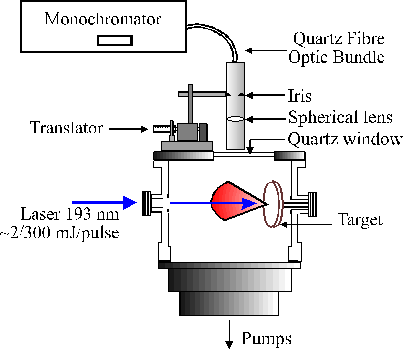 The other end of the fibre bundle butts up against the entrance slit of either of two monochromators. One is a 12.5 cm monochromator (Oriel), equipped with a 600 lines/mm ruled grating and an Instaspec IV CCD array detector, which is capable of providing low resolution dispersed emission spectra covering a wide (~300 nm) range of wavelengths from just a single laser shot, the signal to noise of which can be enhanced by summing for many laser shots.
The second is a 0.5 m Spex 1870 monochromator equipped with a 2400 lines/mm holographic grating and entrance and exit slits whose widths are user selectable. Light emerging from the exit slit of this monochromator is detected with a red sensitive photomultiplier tube (PMT). Higher resolution emission spectra were obtained by scanning the monochromator with the slit widths set narrow and passing the PMT output via a boxcar and a V/F converter to a PC.
Spectra
Back to :-
![]() Bristol University Chemistry Home Page.
Bristol University Chemistry Home Page.